Ewing Sarcoma

Demographic and clinical details: 24-year-old pregnant patient, admitted with soft tissue mass in anterior abdominal wall.
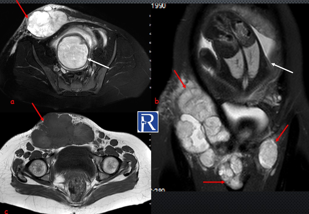
Image Details: Axial (a) and coronal (b)T2 Fat saturated and axial T1 weighted (c) images shows the huge well circumscribed soft tissue mass (red arrows) with heterogeneous signal intensity, located anterior to the right rectus abdominus muscle. The lesion which extends from the anterior superior iliac spine level to the level of symphysis pubis is 15 cm in maximum size. Triple sign which classically described in synovial sarcoma is seen within the mass on T2 W images (a, b). Fetal head (a) and body(b) are seen also (white arrows). Radiological findings (size greater than 5 cm, heterogeneous signal intensity of lesion on T2 and T1 W images) suggest that the lesion is consistent with the malignant lesion. The pathological diagnosis was consistent with soft tissue Ewing's sarcoma.
Soft tissue Ewing sarcoma (STES) is included in the undifferentiated small round cell sarcomas of bone and soft tissue in 2020 WHO classification. Small round cell tumors are a molecularly heterogeneous group lesions characterized by primitive round cell proliferation. When compared with Ewing sarcoma of bone, STES is rare. It is generally found in younger patients (average age is 20 years of age.
Triple sign which is a mixture of high, intermediate and low signal regions within the mass seen on T2 weighted images is seen in our case. It is classically described in synovial sarcoma which is generally found also in younger patients.
In my experience, soft tissue mass lesions with heterogenous signal intensity on T1 and T2 W images should be carefully evaluated for the possibility of malignancy


0 COMMENTS
These issues are no comments yet. Write the first comment...