Peripheral Neurofibroma
Demographic and clinical details: 32 years old female, admitted with soreness and tingling sensation in her left leg.
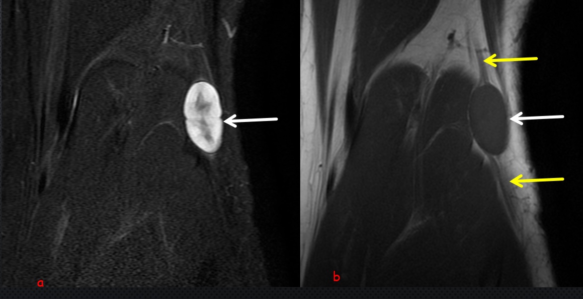
Image Details: Coronal STIR (a) and T1W images shows the typical fusiform shaped soft tissue mass (white arrow) with tapered ends that arise from the peroneal nerve at the level of posterolateral part of knee joint. Note target sign (a) which is characterized by peripheral hypertense signal surrounding the central hypointense signal. Note also the string sign (yellow arrow) (b)which refers to the peroneal nerve entering proximally and exiting distally. Radiological findings suggest the diagnosis of peroneal neurofibroma
Neurofibromas are benign nerve tumors that arise within nerve fascicles. The target sign (a)which is caused by the zonal architecture of the lesion with more myxoid material peripherally and more fibrous tissue centrally was initially believed to be pathognomonic of neurofibroma, it has been observed in both neurofibromas and schwannomas.
In my experience MRI signal changes and lesion shape are important for the diagnosis of neurofibroma. Location of lesion along the course of peripheral nerve is also important to establish the diagnosis.

0 COMMENTS
These issues are no comments yet. Write the first comment...