Sacroillitis

Demographic and clinical details: 32-year-old male patient was admitted with low back pain for 5 years.
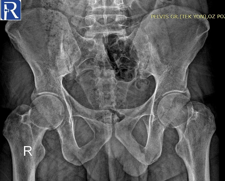
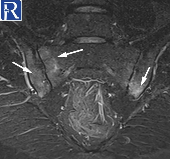
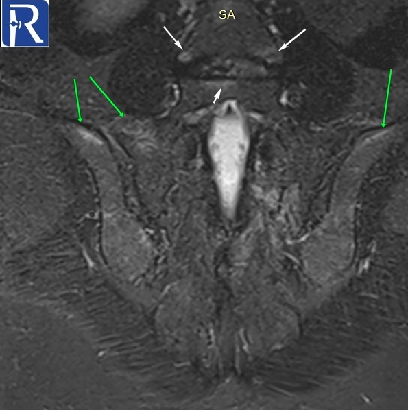
Image Details: AP pelvis X-ray shows the joint space irregularity and peri-articular sclerosis of bilateral and narrowing of the superior part of the left sacrooiliac joints. There are mltiple erosions in both sides. Findings are consistent with radiographic sacroiliitis according to the New York criteria (Grade II sacroiliitis of right, grade III sacroiliitis of left side). Structural lesions indicated with arrows (fatty metaplasia with white arrows, backfill with the green arrow, erosion with black arrow) are seen on T1 W images. Extensive edema of Active sacroiliitis (Extensive bone marrow edema of osteitis (white arrows) is seen on the STIR image. Findings are consistent with active enthesitis (green arrows) and spondylitis (thin white arrows) are also seen on subsequent coronal STIR images. Active arthritis of left L5-S1 facet joint (blue arrow) and active enthesitis of posterior part of the left iliac bone (green arrow) are seen on the fat-suppressed T2 W image. Radiological findings are consistent with radiographic axial spondyloarthritis (ankylosing spondyloarthritis).

.jpg)


.jpg)


0 COMMENTS
These issues are no comments yet. Write the first comment...