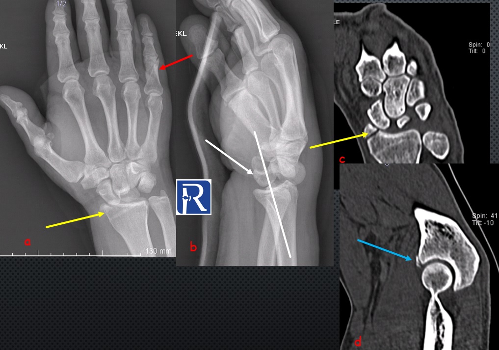
Chauffeur fracture with perilunate dislocation

Demographic and clinical details: 55 years old male, admitted with falling on loutstretched hand
Image Details; AP radiograph of left elbow (a) shows fracture of radial styloid (yellow arrow) and 5th proximal phalanx. Lateral radiograph (b) demonstrates posterior dislocation of capitate, consistent with perilunate dislocation. Axial CT image (c) shows the radial styloid fracture. Sagittal CT image (d) shows the coronoid fracture of ulna (blue arrow).
Distal radius fractures are the most common orthopaedic injury and generally result from fall on an outstretched hand. Intra-articular fractures of the radial styloid process is known as Chauffeur fracture. AP radiography is better than lateral radiography for demonstration of this fracture.
Perilunate dislocation involve dislocation of the carpal bones relative to the lunate which remains in normal alignment with the distal radius. These dislocation may be missed if Radius-lunate-capitat alignment is not properly evaluated on lateral radiographs. Capitate bone dislocated posteriorly in most of the cases.
Ulna coronoid process fractures are rareand often occur in association with elbow dislocation. CT is generally necessary for fracture characterization.
Fractures of hand, elbow, forearm, elbow, humerus and shoulder can be seen in patients who admitted with a fall on outstrcthed hand.


0 COMMENTS
These issues are no comments yet. Write the first comment...