Demographical and clinical details: 53 years old male, tender mass of left-hand that develops 2 months after woody foreign body penetrating injury. ...
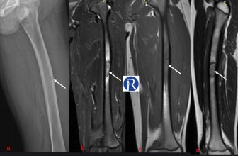
Demographical and clinical details: 9 years old male child, admitted with left thigh pain Image Details: Oblique-lateral femur X-Ray (a) shows the ...
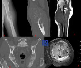
Demographic and clinical details: 64-year-old, male patient, admitted with swelling and pain in the left arm- Image Details: Humerus frontal X-ray ...
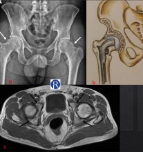
Demographic and clinical details: 32 years old male, with a bilateral hip pain Image Details; AP radiograph of pelvis shows asphericity of the bila ...
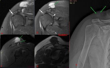
Demographic and clinical details: 73 years, Female, pain and soft tissue mass in right shoulder Image Details: Coronal (a,b), Sagittal (c) fa ...
Image Detail: Sequential Axial plane T1W images demonstrates a triangular shaped accessory muscle (red star) abuts the adjacent neurovascular bundle ( ...
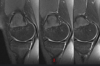
Demographic and clinical details: 15 years old male who had trauma history 3 months ago admitted with right knee pain. İmage Detail: Consecutive sa ...
Image Details. Xray’s and MRI s in 1st figure shows the enchondroma (EC), atypical cartilaginous tumor (ACT) and High-grade chondrosarcoma (CS). ...
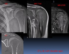
Radiography (DR) is the main diagnostic method of bone tumors. Aggressive Permeative lytic lesion is seen in proximal humerus on DR. Pathological frac ...


