Isolated Lesser Trochanter Avulsion in Adults: A Radiologic Clue to Metastatic Disease

Clinical Presentation
A 75-year-old male with a known history of prostate carcinoma presented with right hip pain and an antalgic gait. He denied any trauma or prior orthopedic intervention. Physical examination revealed pain with hip movement but no palpable mass or deformity.
Imaging Findings
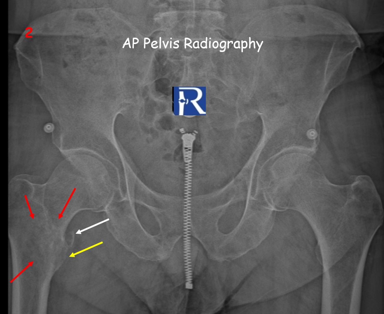
Plain radiography (Figures 1–2) demonstrated a displaced avulsion fracture of the right lesser trochanter with a cortical bone defect and a free bone fragment along the medial aspect of the femoral neck. Additionally, a permeative lytic pattern was observed in the intertrochanteric region.
CT imaging (Figures 3–4) confirmed the avulsion fracture and better delineated the underlying osteolytic lesion at the lesser trochanter, consistent with cortical destruction.
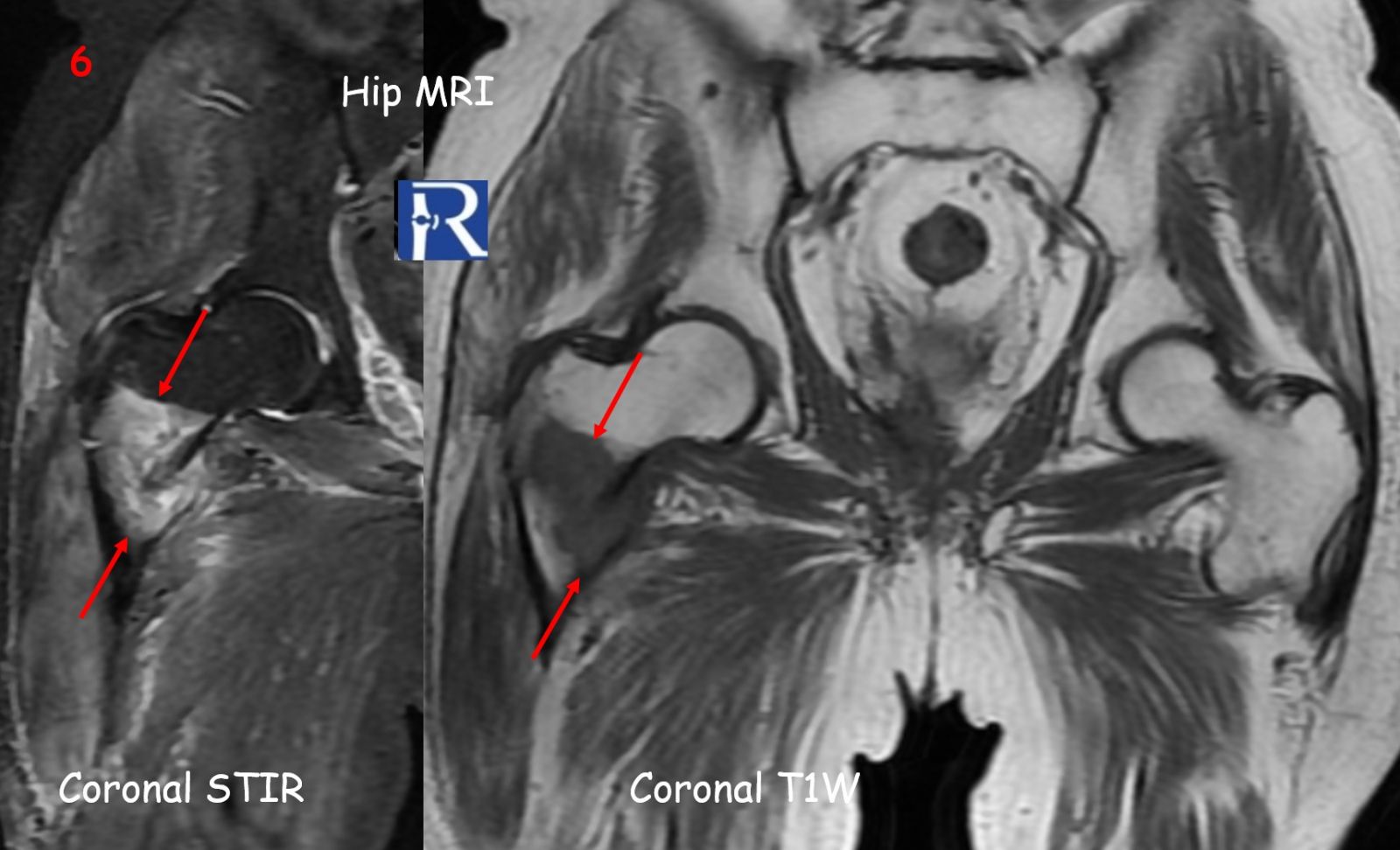
MRI (Figures 5–6) performed two weeks later revealed progression to a pathologic fracture and extension of a hypointense soft-tissue lesion into the intertrochanteric area, compatible with metastatic infiltration.
Figure 7 shows a comparative view between this adult metastatic avulsion fracture and a traumatic apophyseal avulsion of the lesser trochanter in a 16-year-old adolescent.
Figure Legend:
⚪ White arrows → Avulsed bone fragment
???? Yellow arrows → Cortical defect at the lesser trochanter
???? Red arrows → Tumoral infiltration margins
Discussion
Isolated avulsion fracture of the lesser trochanter is rare in adults. In contrast to adolescents, where it is typically caused by sudden and forceful iliopsoas contraction during sports activity, non-traumatic avulsion in adults should always raise suspicion for an underlying pathological process.
In most cases, the etiology is metastatic, particularly from prostate, lung, colon, and thyroid carcinomas. In the literature, approximately 70% of reported adult cases were associated with metastatic disease.
Imaging plays a crucial role in diagnosis and management:
- Radiographs often provide the initial clue but may underestimate soft-tissue involvement.
- MRI is highly sensitive for defining the extent of marrow and soft-tissue infiltration and for surgical planning.
- CT aids in evaluating cortical destruction and can guide biopsy when necessary.
Prompt recognition of this radiologic pattern is vital, as the finding may represent the first manifestation of an occult malignancy.
Teaching Points
- In adults, an isolated lesser trochanter fracture without trauma is almost always pathologic.
- Metastatic disease should be the first consideration in differential diagnosis.
- MRI is essential for evaluating the full extent of infiltration and distinguishing between metastasis and primary bone tumors.
- Early recognition of this subtle radiographic sign can lead to timely diagnosis of an underlying malignancy, often before systemic symptoms appear.


0 COMMENTS
These issues are no comments yet. Write the first comment...